Light Microscope
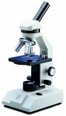
Light microscope were first used by the Renissance and were first invented in the 1590's. Light microscopes let visible light passes through the specimen and glass lenses. The lenses refracts light into the eye piece on the microscope. Microscope maginifes and resolute objects that are very small into a visible size that human eyes can not see. Magnification/maginify is the ratio of an object image to it's real size and resolution/resolute is the clarity of the image. Light microscopes cannot resolve images/details that are finer than .2 micrometer or 200 nanometer.
Electron Microscope
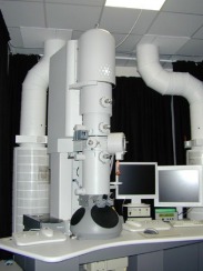
Cell were first discovered by Robert Hooke but light microscopes can not see details of the cell so Electron Microscopes were invented in the 1590's that focus on the surface and the details within cells. There are two types of electron microscopes: Transmission electron microscope (TEM) and Scanning electron microscope(SEM). Transmission electron microscope are used to focus on the interal untrastructure of cells. Scanning electron microscope focus on the surface of the cell. A disadvantage of electron microscope is that it give fine detail of the cell and it's organelle but some specimen may kill the cells.
Cell Fractionation
Cell fractionaiton is the separation of organelles from it cells'. The instrument uses centrifuge that spin test tubes in a various speed. The most powerful machine that is used in this process is called ultracentrifuge.
